

VitroGel® 3D High Concentration
Overview
VitroGel® 3D is a pure and unmodified hydrogel that allows the maximum flexibility to manipulate the 3D cell culture environment for different needs. The unmodified hydrogel matrix structure is good for cell spheroid formation, suspension cells or cells requiring low cell-matrix interactions.
VitroGel 3D is one system of the family of ready-to-use, xeno-free tunable hydrogel system which closely mimics the natural extracellular matrix (ECM) environment.
VitroGel creates a functional and optimized environment to make cells feel like at home. The hydrogel system is room temperature stable, has a neutral pH, transparent, permeable and compatible with different imaging systems. The solution transforms into a tunable hydrogel matrix by simply mixing with the cell culture medium. Cells cultured in this system can be easily harvested out with our VitroGel Cell Recovery Solution. The hydrogel can also be tuned to be injectable for in vivo studies.
From 3D cell culture, 2D cell coating to animal injection, VitroGel makes it possible to bridge the in vitroand in vivo studies with the same platform system.
Specifications
| Contents | VitroGel® 3D High Concentration, 3 mL VitroGel® Dilution Solution, 50 mL |
| Hydrogel Formulation | Xeno-free tunable hydrogel, pure and unmodified. HIGH CONCENTRATION |
| Use | Good for cell spheroid formation, suspension cells or cells require low cell-matrix interactions |
| Operation | Room temperature |
| Hydrogel Strength | 10 to 4,000 Pa of G’ depending on dilution ratio. Dilute with VitroGel Dilution Solution (TYPE 1 or TYPE 2) for different concentrations. |
| pH | Neutral |
| Color | Transparent |
| Cell Harvesting | 20 min cell recovery using VitroGel Cell Recovery Solution |
| Injectable | Injectable hydrogel |
| Storage | Store at 2-8°C. Ships at ambient temperature |
Product Documentation
Data
-
3D Cell Culture Applications
-
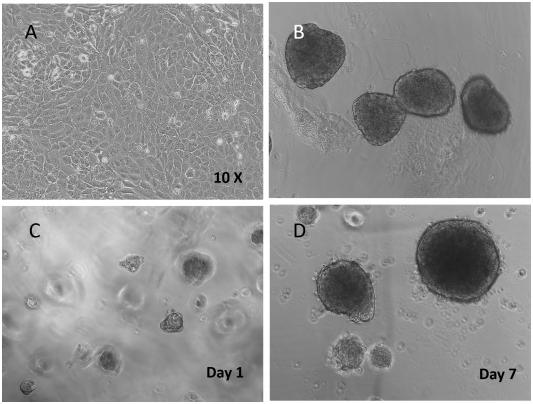
Figure 1. Beta Lox 5 (BL5) cells 3D culture in VitroGel 3D system.
A. BL5 cells culture on the surface of regular tissue culture treated well plate (control); B. Normal human islets grew in suspension culture (comparison); C. 3D culture of BL5 cells in VitroGel 3D at Day 1; D. 3D culture of BL5 cells in VitroGel 3D at Day 7. Under 3D culture of VitroGel 3D, BL5 cells form islet-like structures very similar to normal human islets. The hydrogel is prepared at 1:3 dilution. The images were taken at 10X magnification.

-
Figure 2. CD8+ T cells 3D culture in VitroGel 3D system
CD8+ T cells culture grew in suspension culture (contorl); B. 3D culture of CD8+ T cells in VitroGel 3D at Day 7. CD8+ T cells are vibrant in 3D culture conditions of VitroGel 3D. The cells can easily move within the unmodified hydrogel matrix. The hydrogel is prepare at 1:3 dilution. The images were taken at 10X magnification.
-
2D Coating Applications
-
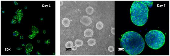
Figure 3. Human colon cancer cells (HCT 116) cells cultured on top of VitroGel 3D hydrogel
A thick hydrogel coating plate has been prepared by mixing VitroGel 3D with PBS at 1:1 ratio. A 300 µL mixture has been added to a well of a 24-well plate and stabilization at room temperature for 20 min before adding cells on top of the hydrogel. Cell spheroids form on the top of the hydrogel. Cells seeded at 2.5-10×105 cells/mL.
-
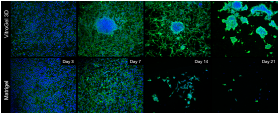
Figure 4. Comparison of long-term neuronal culture seeded onto thick hydrogel mats.
Cells are stained with Beta-III-Tubulin (green) cytoskeleton marker and their nuclei are counter-stained with DAPI (blue). Cells spread out and form neural-like networks as early as day 3 post-differentiation, with comparable efficacy between VitroGel 3D and Matrigel, based on cell survival, culture spreading and morphological analysis reached between days 7 and 9. On Matrigel mats, cell culture health and viability drops off sharply once day 9 has passed, with most cells detaching and neurites retracting by day 14 and the vast majority of cells gone by day 21. If grown onto VitroGel 3D mats, differentiated B35 neurons have a tendency to self-organize into 3D clusters very early on (Day 7), assuming a mixed 2D/3D cell culture for the first two weeks of the time-course. By Day 21, these cells have migrated into self-assembled 3D clusters, embedded into the thick hydrogel matrix, with very few cells between the clusters, but without any significant cell death.
-
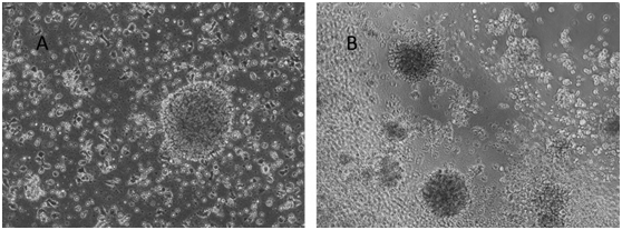
Figure 5. Human Lymphoblastoid Priess cells cultured on top of VitroGel 3D hydrogel
A. Priess cells grown in suspension (control); B. Priess cells grown on top of VitroGel 3D at day 7. A hydrogel substance can be prepared with different stiffness by adjusting the dilution of VitroGel 3D from 1:1 to 1:3 ratio. Cells seeded on the top of the hydrogel form cell spheroids form on the top of hydrogel. The hydrogel provides a soft substance for cell to attach and grow.